

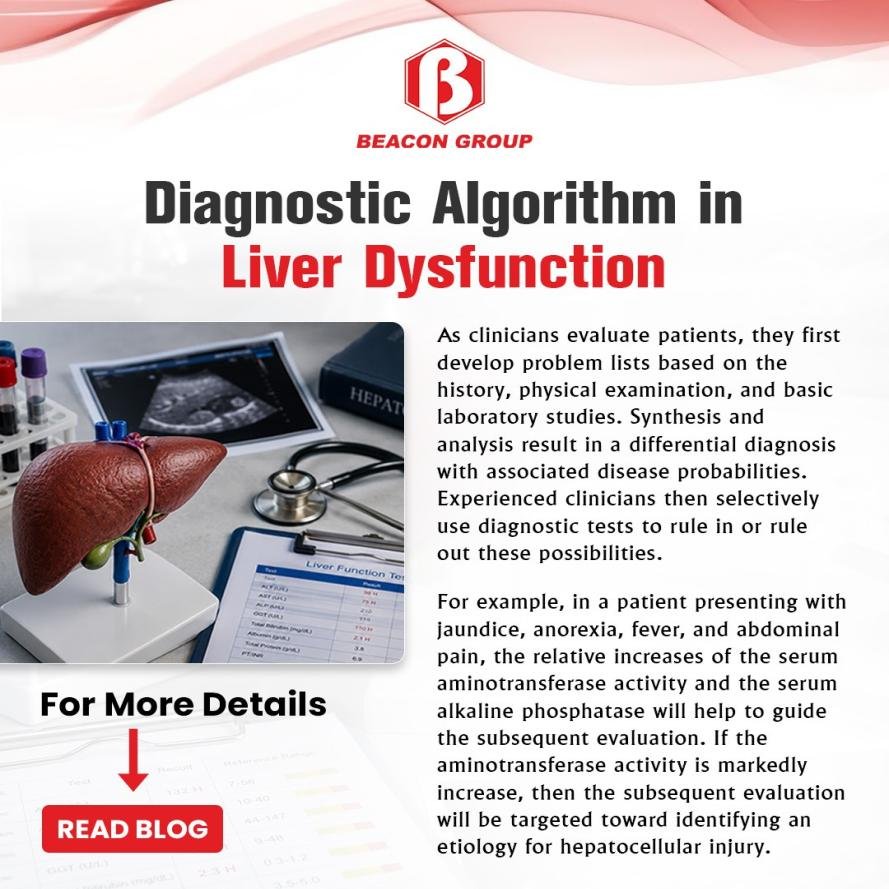
As clinicians evaluate patients, they first develop problem lists based on the history, physical examination, and basic laboratory studies. Synthesis and analysis result in a differential diagnosis with associated disease probabilities. Experienced clinicians then selectively use diagnostic tests to rule in or rule out these possibilities. For example, in a patient presenting with jaundice, anorexia, fever, and abdominal pain, the relative increases of the serum aminotransferase activity and the serum alkaline phosphatase will help to guide the subsequent evaluation. If the aminotransferase activity is markedly increase, then the subsequent evaluation will be targeted toward identifying an etiology for hepatocellular injury. In contrast, if the alkaline phosphatase is markedly increased, then the evaluation would be targeted toward identifying an etiology for obstructive jaundice.
The clinical diagnostic algorithm for evaluating liver disease relies on initial laboratory testing, pattern recognition of enzyme elevations, non-invasive imaging or fibrosis scoring, and targeted etiology screens.
Initial blood panel: Run alanine aminotransferase (ALT), aspartate aminotransferase (AST), alkaline phosphatase (ALP), gamma-glutamyl transferase (GGT), total bilirubin, and albumin.
Hepatocellular pattern: Markedly elevated ALT/AST points to cell injury from viral hepatitis, medications, or metabolic dysfunction-associated steatotic liver disease (MASLD).
Cholestatic pattern: Markedly elevated ALP and GGT points to bile duct obstruction or infiltrative disease.
Viral screening: Test for Hepatitis B surface antigen and Hepatitis C antibody.
Metabolic/Toxic workup: Assess iron saturation, fasting glucose/lipids, complete alcohol history, and review current medications or supplements.
Autoimmune markers: Order autoimmune panels (such as ANA, ASMA) if primary screens are negative and enzymes remain high. Imaging and Fibrosis Staging.
Abdominal ultrasound: Perform first-line imaging to check for steatosis, biliary dilation, or structural masses.
Non-invasive elastography: Use transient elastography (FibroScan) to quantify liver stiffness and stage fibrosis.

Beacon Group
424, New GIDC, Kabilpore, Navsari, Gujarat 396424 India

Beacon Diagnostics Europe Srl.
Via Aliprandi 41, 20851, Lissone(MB), Italy

Beacon Diagnostics - Spain
C/Sicilia 159, 08013, Barcelona, Spain

Beacon Diagnostics-Philippines
Unit 108 Two Premier 1st St. Alabang Premier, Alabang Zapote Rd, Concha Cruz Drive, Almanza Uno, Las Pinas City